DD3 — Arms, Wrists & Hands
The 27 Bones of Your Hand, the Ulnar Nerve Trap, and Why Your Grip Pressure Matters
📋 DOCUMENT MAP
This DD covers the entire arm chain from shoulder to fingertip: humerus, elbow (cubital tunnel — where the ulnar nerve gets trapped), forearm muscles, the 27 bones of the hand, the 8 carpal bones in 2 rows, and the 9 tendons + median nerve of the carpal tunnel.
Why this matters for a 50+ tennis player: the elbow and wrist are where most recreational tennis injuries appear — tennis elbow (lateral epicondylitis), cubital tunnel syndrome, carpal tunnel syndrome, De Quervain's tenosynovitis. Most are caused by grip pressure that's too high, too low, or held too long.
What it does NOT cover: the shoulder (DD2), the cervical spine nerves that also feed the arm (DD4 Trunk & Spine), or stroke mechanics (Forehand/Backhand deep dives).
Reading time: 35–45 minutes.
📑 TABLE OF CONTENTS
| # | English |
|---|---|
| 1 | The Humerus, Radius, Ulna — Your Arm's 3 Bones |
| 2 | The Cubital Tunnel — The #1 Trap for Tennis Players |
| 3 | The Ulnar Nerve Pathway — From Neck to Pinky |
| 4 | STOP STRETCHING — The Nerve Flossing Solution |
| 5 | The 27 Bones of the Hand — A 3-Zone Machine |
| 6 | The Carpal Tunnel — Where 9 Tendons + 1 Nerve Live |
| 7 | Grip Pressure — The 3/10 to 7/10 Rule |
| 8 | Tendon Gliding — Daily Maintenance for Tennis Hands |
| 9 | The ECRB and the Mechanical Side of Tennis Elbow |
Chapter 1 — The Humerus, Radius, Ulna (Your Arm's 3 Bones)
The arm is 3 bones connected by 3 joints. Upper arm = humerus (single bone). Forearm = radius (thumb side) and ulna (pinky side). The joints are the shoulder, elbow, and wrist.
The elbow is a hinge joint. Two movements only — flexion (bending) and extension (straightening). It cannot rotate. The rotation you feel at the elbow is actually the RADIUS rotating around the ULNA in the forearm (called pronation/supination).
The 3 Bones — Where They Meet
| Bone | Location | Where It Meets Others | Tennis Role |
|---|---|---|---|
| Humerus | Upper arm, single long bone | Shoulder (with scapula), elbow (with radius + ulna) | Carries the elbow flexors (biceps, brachialis) and extensor (triceps). |
| Radius | Forearm, THUMB side | Elbow (with humerus), wrist (with scaphoid + lunate) | Rotates around ulna for pronation/supination. Forehand supination is critical. |
| Ulna | Forearm, PINKY side | Elbow (with humerus via olecranon), wrist (with triquetrum) | The "fixed" bone of the forearm. The olecranon forms the elbow tip. The ulnar nerve runs behind it. |
The 3 Joints — Where They Move
| Joint | Type | Movement Range | Tennis Translation |
|---|---|---|---|
| Elbow | Hinge | 0° (extended) to ~145° (flexed) | Most strokes need 90–110°. Biceps curl = full flexion. Locked-out triceps = 0°. |
| Radioulnar (proximal + distal) | Pivot | ~150° pronation-supination | Forehand supination (palm up at contact) requires ~80–90° supination. Backhand slice uses ~30° pronation. |
| Wrist | Ellipsoid (modified ball-and-socket) | Flexion ~80°, extension ~70°, radial deviation ~20°, ulnar deviation ~30° | The "fine adjustment" joint. The 70/30 rhythm of forehand is wrist-driven. |
The Forearm Muscles — 20+ Muscles, 4 Functions
| Function | Primary Muscles | Tennis Role |
|---|---|---|
| Elbow flexion | Biceps brachii, brachialis, brachioradialis | The "lift" of the racquet during backswing. Brachialis is the true workhorse — biceps adds supination. |
| Elbow extension | Triceps brachii (3 heads), anconeus | The "push" through contact. Triceps fires concentrically in last 0.1 sec before ball leaves racquet. |
| Pronation (palm down) | Pronator teres, pronator quadratus | Topspin forehand uses 20–40° pronation during forward swing. |
| Supination (palm up) | Biceps brachii, supinator | Forehand volley and slice use full supination at contact. |
| Wrist flexion | Flexor carpi radialis, flexor carpi ulnaris, palmaris longus | The "lag" position before forward swing. Loaded flexors release during contact for whip. |
| Wrist extension | Extensor carpi radialis longus + brevis, extensor carpi ulnaris | The "layback" position in backswing. Stretched extensors add 5–10% racquet head speed. |
| Wrist radial deviation | Extensor carpi radialis longus + brevis, flexor carpi radialis | Topspin forehand uses ~10° radial deviation at contact. |
| Wrist ulnar deviation | Extensor carpi ulnaris, flexor carpi ulnaris | Backhand uses ~15–20° ulnar deviation at contact. |
Source: Roetert & Kovacs, Tennis Anatomy, Ch.3 pages 75–79.
Chapter 2 — The Cubital Tunnel (The #1 Trap for Tennis Players)
The cubital tunnel is a groove behind the medial epicondyle of the humerus (the "funny bone" knob on the inside of your elbow). The ulnar nerve passes through it, held down by a ligament called Osborne's ligament. When the elbow bends, the tunnel NARROWS by 55%. Pressure inside the tunnel spikes.
This is the most common nerve compression site in the elbow. Symptoms: tingling in the ring + pinky fingers, weakness in grip, "falling asleep" feeling when holding the phone. In tennis players, the one-handed backhand is the #1 cause — the elbow bends past 90° for hundreds of strokes.
The Cubital Tunnel Numbers
| Number | What It Means | Tennis Implication |
|---|---|---|
| 55% | Tunnel cross-section reduction when elbow flexes from 0° to 90° | One-handed backhand with elbow bent 90°+ = sustained 55% compression for entire stroke. |
| +50% | Increase in intraneural pressure at 90° flexion | The nerve is literally being squeezed. Over hundreds of strokes → ischemia → nerve damage. |
| 7× | Repetition multiplier for backhand (vs other strokes) | 50 forehands vs 50 backhands: the backhand compresses the nerve 7× more cumulatively. |
| 2 compression sites | Cubital tunnel AND Guyon's canal (wrist) | Ulnar nerve has TWO compression sites on the path to the pinky. Either can fail. |
The Double Crush Phenomenon
The ulnar nerve can be compressed at the C8-T1 nerve root (neck), at the cubital tunnel (elbow), AND at Guyon's canal (wrist). If even one site is mildly compressed, the nerve becomes sensitive. A second compression site — even a small one — produces symptoms out of proportion.
Tennis truth: most recreational players with persistent "tennis elbow" actually have DOUBLE crush — neck compression from poor posture (DD4) PLUS cubital tunnel compression from one-handed backhand. Treating only the elbow misses the neck.
The diagnostic: a neurologist can map the compression site with EMG/nerve conduction studies. If you have persistent symptoms despite 6 weeks of tennis-elbow treatment, ASK for nerve testing.
Source: Anatomy_Tay_Than_Kinh_Full.docx, Part I. Tennis Anatomy Ch.10 (Common Injuries) corroborates with cubital tunnel syndrome details.
Chapter 3 — The Ulnar Nerve Pathway (From Neck to Pinky)
The ulnar nerve starts at C8-T1 nerve roots in your lower neck, travels down the arm, passes BEHIND the medial epicondyle at the elbow (the cubital tunnel), then continues through Guyon's canal at the wrist, and ends in the ring and pinky fingers. It is the longest unprotected nerve in the arm.
Its job: controls the flexor carpi ulnaris (wrist flexor on pinky side), the flexor digitorum profundus to ring + pinky, the intrinsic hand muscles (interossei, lumbricals, hypothenar), and provides sensation to ring + pinky + ulnar half of palm.
When it fails: "claw hand" deformity (ring + pinky can't flex at MCP), wasting of the hypothenar eminence, weakness in grip (because FDP to last 2 fingers is paralyzed). Tennis player can't hold the racquet.
The Ulnar Nerve Path — 5 Stops
| Stop | Location | What Happens | Tennis Translation |
|---|---|---|---|
| 1 | C8-T1 nerve roots (lower neck) | Origin | Compression here = neck posture issue. See DD4. |
| 2 | Thoracic outlet (between collar bone and 1st rib) | Tight space near scalenes | Rare tennis site. More common in swimmers. |
| 3 | Cubital tunnel (behind medial epicondyle at elbow) | The #1 trap. Tunnel narrows 55% at 90° flexion | One-handed backhand with elbow bent. |
| 4 | Forearm (between FCU muscle heads) | Less common compression | Repetitive gripping. |
| 5 | Guyon's canal (at wrist, between pisiform and hamate) | The #2 trap. Carpal-bone-defined tunnel | Grip pressure when wrist is ulnar-deviated. |
The 5 Warning Signs (When to See a Neurologist)
| Warning Sign | What It Means | Action |
|---|---|---|
| 1. Tingling in ring + pinky lasting >5 min after play | Acute nerve irritation | Reduce backhand volume, nerve floss 3×/day |
| 2. Dropping the racquet during play | Motor weakness — FDP failing | STOP PLAY. See neurologist. EMG/nerve conduction. |
| 3. Wasting of the hypothenar eminence | Visible hollow between pinky and wrist | URGENT neurology referral. Chronic compression. |
| 4. Claw hand posture at rest | Intrinsic muscle paralysis | URGENT. May need surgical decompression. |
| 5. Numbness that doesn't resolve in 24 hours | Persistent nerve ischemia | See neurologist within 48 hours. |
Source: Anatomy_Tay_Than_Kinh_Full.docx, Part I, paragraphs 4-12. Tennis Anatomy Ch.10 corroborates.
Chapter 4 — STOP STRETCHING (The Nerve Flossing Solution)
The most common advice for "tennis elbow" is WRONG: stretch the forearm, stretch the wrist, stretch the elbow. But the ulnar nerve doesn't want to be STRETCHED. Stretching increases intraneural strain >15%. This reduces blood flow inside the nerve (microvascular ischemia).
The fix is NERVE FLOSSING — also called nerve gliding. The nerve slides back and forth between its endpoints. You flex the wrist while extending the elbow, then extend the wrist while flexing the elbow. This creates motion at the nerve WITHOUT stretching it.
The Nerve Flossing Technique — Step by Step
| Step | Position | Hold | Why |
|---|---|---|---|
| 1. Start | Elbow extended, wrist flexed (bent down), shoulder depressed (down) | 2 sec | Long nerve position. |
| 2. Slide | Slowly: elbow flexes (bend), wrist extends (bent up), shoulder elevated (up) | 2 sec | Nerve slides "up" toward the head. |
| 3. Return | Slowly: elbow extends, wrist flexes, shoulder depressed | 2 sec | Nerve slides "down" toward the fingers. |
| 4. Repeat | 10 slow reps, 3× per day | Before and after play | Restores nerve mobility, reduces intraneural pressure. |
The "Pain Cap" Rule — Keep It Under 3/10
Nerve flossing should HURT LESS than 3/10 on a 0–10 scale. If you feel tingling, electric shocks, or pain >3/10, STOP. You are compressing or over-stretching the nerve. The goal is gentle motion, not aggressive stretching.
The 50+ protocol: 10 slow reps in the morning (before the day stiffens the nerve), 10 reps mid-day (counteract the desk posture), 10 reps after tennis (clear inflammation). Total: 30 reps/day, 3 minutes.
The 4 Common Mistakes With Nerve Flossing
| # | Mistake | Why It's Wrong | Fix |
|---|---|---|---|
| 1 | Doing it too fast | Fast motion = stretch, not glide | Slow. 1 rep every 3 seconds. |
| 2 | Pulling hard on the head or fingers | This stretches the nerve | Move only at the wrist + elbow. Head and shoulders RELAXED. |
| 3 | Doing it when the nerve is acutely inflamed (>5/10 pain) | Flare-up risk | Wait until pain drops below 3/10. Use ice first. |
| 4 | Only doing it after symptoms appear | Reactive, not preventive | Make it a daily habit, like brushing teeth. |
Source: Anatomy_Tay_Than_Kinh_Full.docx, Part I, paragraph 10-14. Tennis Anatomy Ch.10 corroborates.
Chapter 5 — The 27 Bones of the Hand (A 3-Zone Machine)
The human hand has 27 bones, 27 joints, 34 muscles (intrinsic + extrinsic), over 100 ligaments, and 3 main nerves (median, ulnar, radial). It is the most complex mechanical structure in the body. The hand is divided into 3 zones: carpals (wrist), metacarpals (palm), phalanges (fingers + thumb).
The 3 Zones of the Hand
| Zone | # Bones | Names | Tennis Role |
|---|---|---|---|
| Carpals (wrist) | 8 | Scaphoid, lunate, triquetrum, pisiform (proximal row); trapezium, trapezoid, capitate, hamate (distal row) | The wrist platform. Must absorb racquet vibration without damage. The "carpal boss" is a common tennis injury at the index CMC. |
| Metacarpals (palm) | 5 | Metacarpals I–V (thumb to pinky) | Form the palm. The 2nd and 3rd metacarpals are FIXED (immobile) — they form the rigid platform of the hand. |
| Phalanges (fingers + thumb) | 14 | 2 in thumb (proximal + distal), 3 in each finger (proximal + middle + distal) | The grippers. Each finger has 3 joints (MCP, PIP, DIP) except thumb (2 joints: MCP, IP). |
The 8 Carpal Bones in 2 Rows — A Map
| Row | Bones (lateral → medial) | Function |
|---|---|---|
| Proximal (closer to forearm) | Scaphoid, Lunate, Triquetrum, Pisiform | Articulate with radius. Form the wrist joint proper. Most wrist fractures happen here. |
| Distal (closer to fingers) | Trapezium, Trapezoid, Capitate, Hamate | Articulate with metacarpals. The hook of hamate + pisiform form Guyon's canal (ulnar nerve). |
The Thumb — 50% of Hand Function
The thumb accounts for 50% of total hand function. This is because of OPPOSITION — the ability to bring the thumb pad across to meet the pads of the other 4 fingers. No other finger can do this. Loss of opposition = loss of grip, loss of fine motor.
Tennis implication: the thumb's role in grip pressure is HUGE. In the Eastern backhand grip, the thumb sits on bevel 7 of the handle. It acts as a counterforce against the fingers. If the thumb is over-gripping (the common 3.5 mistake), the ulnar nerve gets squeezed at Guyon's canal.
De Quervain's Tenosynovitis: inflammation of the tendons APL (abductor pollicis longus) and EPB (extensor pollicis brevis). These pass through the 1st compartment of the wrist. Common in tennis players who grip with the thumb. Pain at the thumb base + radial wrist.
Source: Anatomy_Tay_Than_Kinh_Full.docx, Part II paragraphs 16-26. Tennis Anatomy Ch.3 (Arms and Wrists) and Ch.10 corroborate.
Chapter 6 — The Carpal Tunnel (Where 9 Tendons + 1 Nerve Live)
The carpal tunnel is a narrow passageway on the PALMAR side of the wrist. It's bounded by the carpal bones (floor and walls) and the flexor retinaculum (transverse carpal ligament) as the roof. Inside: 9 flexor tendons + the median nerve. Total cross-section: about 2 cm².
The median nerve controls sensation in thumb, index, middle, and radial half of ring finger. It also controls the thenar muscles (the "flesh pad" at the base of the thumb).
What's Inside the Carpal Tunnel
| # | Structure | Function |
|---|---|---|
| 1–4 | Flexor digitorum superficialis (4 tendons) | Flex PIP joints of fingers 2–5 |
| 5–8 | Flexor digitorum profundus (4 tendons) | Flex DIP joints of fingers 2–5 |
| 9 | Flexor pollicis longus (1 tendon) | Flex thumb IP joint |
| 10 | Median nerve | Sensation to thumb/index/middle/radial ring + thenar motor |
Carpal Tunnel Syndrome — The Trigger
Anything that INCREASES pressure inside the 2 cm² tunnel causes symptoms. The 9 tendons swell with inflammation → takes up more space → median nerve gets squeezed → tingling in thumb/index/middle.
Tennis triggers: (a) sustained tight grip on the racquet, (b) excess wrist flexion during low volleys, (c) excess wrist extension during serves, (d) cold weather (vasoconstriction reduces tunnel space).
The fix: grip pressure 3/10 (see Ch.7). Wrist neutral during volley and serve. Warm up before cold-weather play. Tendon gliding exercises (Ch.8).
The Phalen's Test — A Self-Check
Press the backs of your hands together with wrists fully flexed (fingertips pointing down). Hold 60 seconds. If you feel tingling in thumb/index/middle within 60 seconds, the median nerve is being compressed.
If positive: nerve gliding 3×/day, grip pressure reduction, ergonomic racquet handle (smaller grip size if pain is severe), medical referral if persistent.
Source: Anatomy_Tay_Than_Kinh_Full.docx, Part II paragraphs 18-24.
Chapter 7 — Grip Pressure (The 3/10 to 7/10 Rule)
The 3/10 to 7/10 rule is the single most important hand technique in tennis. Grip at 3/10 while waiting for the ball (relaxed, just enough to keep the racquet from falling). Grip at 7/10 at the moment of contact (firm, but not white-knuckled). Release back to 3/10 in the follow-through.
The amateur mistake: grip at 8–9/10 the ENTIRE point — from ready position to follow-through. This is the primary cause of (a) tennis elbow, (b) carpal tunnel, (c) cubital tunnel syndrome, (d) forearm fatigue. The grip should be a SPEED CHANGER, not a constant force.
The Pressure Numbers — What They Mean
| Pressure | Sensation | What You're Doing | Risk |
|---|---|---|---|
| 1–2/10 | Racquet about to fall | Too loose. No control. | Shanked shots. |
| 3/10 | Hold a tube of toothpaste without squeezing | The "ready" pressure. Energy conserved. | None. |
| 5/10 | Hold a full coffee cup | Mid-range. OK during slow balls. | Cumulative fatigue if held long. |
| 7/10 | Squeeze someone's hand firmly | The "contact" pressure. Maximum useful. | OK if brief. |
| 8–10/10 | Crush a can / wring a towel | Over-grip. The amateur default. | Tennis elbow, carpal tunnel, cubital tunnel. |
The Timing Cue — When to Grip
| Phase | Pressure | Why |
|---|---|---|
| Ready position | 3/10 | Energy conservation. Fast hands. |
| Split-step → first step | 4/10 | Slight increase to "wake up" the forearm. |
| Unit turn (shoulder rotation) | 5/10 | Racquet is moving with the body. |
| Forward swing start (chest turns) | 6/10 | Begin loading the grip. |
| 0.2 sec before contact | 7/10 | The "squeeze" moment. |
| At contact | 7/10 | Maximum pressure. Brief. |
| 0.2 sec after contact | 5/10 | Release. |
| Follow-through | 3/10 | Back to relaxed. Ready for next ball. |
The 50+ Grip Reality — Tendons Need Recovery
The forearm tendons in a 50+ player have less blood supply than at 25. Tendon healing is blood-supply-dependent. Over-gripping creates micro-tears that heal SLOWER than at 25. The 3/10 baseline matters even more after 50.
The drill: the "ball drop" test. Hold a tennis ball in your non-dominant hand at 3/10 pressure. Walk around for 1 minute. If you feel the need to grip harder, you were probably gripping at 6/10 or higher. Reset to 3/10.
Source: Anatomy_Tay_Than_Kinh_Full.docx, Part III paragraphs 28-32.
Chapter 8 — Tendon Gliding (Daily Maintenance for Tennis Hands)
Tendon gliding exercises move each tendon through its full range of motion. The goal is to (a) prevent adhesions between tendons and their sheaths, (b) maintain synovial fluid flow, (c) detect early signs of inflammation. They take 2 minutes. They are the BEST daily maintenance for tennis hands.
The 5 positions of tendon glide: straight → hook fist → full fist → tabletop → straight. Each position stresses different tendons differently. The full cycle takes 5 seconds per hand.
The 5 Tendon Glide Positions
| Position | Hand Shape | Tendons Stressed | Hold |
|---|---|---|---|
| 1. Straight | Fingers straight, palm down | All tendons in resting length | 1 sec |
| 2. Hook fist | DIP + PIP flexed, MCP straight | FDP (deep flexor) at maximum glide | 1 sec |
| 3. Full fist | All joints flexed | FDS + FDP together | 1 sec |
| 4. Tabletop | MCP flexed 90°, PIP + DIP straight | FDS at maximum glide, lumbricals active | 1 sec |
| 5. Straight (return) | Same as #1 | Synovial fluid redistribution | 1 sec |
The Tennis-Specific Glide Sequence — When to Use It
| When | Sequence | Purpose |
|---|---|---|
| Morning | 10 reps each hand, slow | Restore overnight stiffness |
| Before play | 5 reps each hand, dynamic | Warm tendons, detect inflammation |
| During match (changeover) | 5 reps each hand | Reduce cumulative tendon load |
| After play | 10 reps each hand, slow | Distribute synovial fluid, reduce adhesion |
| If any pain >3/10 | STOP. Ice 10 min. Glide gently after ice. | Don't glide into inflammation |
The 3 Signs You Need More Than Gliding
| Sign | What It Means | Action |
|---|---|---|
| 1. Pain during glide that wasn't there before | Acute inflammation | Ice + rest. See doctor if persists >3 days. |
| 2. A tendon "catches" or locks during glide | Stenosing tenosynovitis (trigger finger) | Stop gliding that tendon. Medical referral. |
| 3. Persistent morning stiffness >30 min | Rheumatoid or chronic inflammation | See rheumatologist. May need medication. |
Source: Anatomy_Tay_Than_Kinh_Full.docx, Part III paragraph 32. Tennis Anatomy Ch.10 corroborates the tendon gliding protocol.
📋 DD3 CARD — Printable
The ulnar nerve has 2 trap sites (cubital tunnel + Guyon's canal) and 27 bones form the hand. Grip pressure is the master switch: 3/10 relaxed, 7/10 at contact. Most tennis-arm injuries come from over-grip.
- Cubital tunnel narrows 55% at 90° elbow flexion
- 27 hand bones / 8 carpals / 14 phalanges / 5 metacarpals
- Carpal tunnel cross-section: 2 cm²
- Grip pressure: 3/10 ready → 7/10 contact → 3/10 follow
- 9 tendons + 1 median nerve inside the carpal tunnel
Gripping at 8–10/10 the entire point instead of modulating between 3/10 (relaxed) and 7/10 (contact). This causes tennis elbow, carpal tunnel syndrome, and cubital tunnel syndrome — the three most common tennis-arm injuries in 50+ players.
- Nerve flossing: 10 slow reps, 3×/day (page 4)
- Tendon gliding: 10 reps/hand, morning + after play
- Ball-drop test: walk with ball at 3/10, reset if you instinctively grip harder.
"Relaxed ready, firm contact, relaxed follow."
Chapter 9 — The ECRB and the Mechanical Side of Tennis Elbow
Everything above on the ulnar nerve and cubital tunnel explains the NEUROLOGICAL cause of elbow pain. But most "tennis elbow" — lateral epicondylitis — isn't a nerve problem at all. It's a tendon problem, and it lives on the OUTSIDE of the elbow, not the inside where the ulnar nerve runs.
Lateral epicondylitis is a condition of the origin of the Extensor Carpi Radialis Brevis (ECRB) — one of the wrist-extensor muscles identified in Chapter 1's forearm-muscle table. It's caused by repetitive microtrauma from eccentric loading, not acute inflammation, which is why it responds poorly to rest alone and well to targeted strengthening.
The "Leading Elbow" Fallacy
A common technical error among recreational players is letting the elbow lead — flexed and positioned ahead of the body at impact, instead of staying integrated with the torso's rotation. This forces the wrist extensors, including the ECRB, to work eccentrically to stabilize the racquet head against the ball's impact force. Do that for thousands of strokes over a season and the ECRB origin accumulates the same kind of collagen damage as any other overuse tendinopathy.
| Parameter | Elite Technique | Recreational Error |
|---|---|---|
| Wrist angle at impact | ~23° extension | ~13° flexion |
| Grip pressure | Relaxed immediately post-impact | Constant "death grip" through the shot |
| Elbow position | Integrated with torso rotation | "Leading" the stroke, isolated from the body |
Notice the wrist-angle numbers land inside the range already described in DD1 — The Player in Motion: 0–20° of extension at contact. A recreational player locked into 13° of flexion at impact isn't just losing racquet-head speed — the ECRB is being asked to eccentrically decelerate the racquet from a mechanically disadvantaged position, stroke after stroke.
The Dual-Position Strengthening Protocol
Effective prevention and rehab for ECRB-based tennis elbow isn't about stretching (see Chapter 4's warning about nerve flossing vs. stretching — the same "don't just stretch it" logic applies here, for a different reason: stretching a healing tendon under active eccentric damage doesn't address the loading problem that caused it). It's a two-position strengthening program:
- 90° elbow flexion — isolates the ECRB directly, targeting the muscle belly and origin under a shortened lever arm.
- 180° elbow extension — stretches the extensor group under load, which promotes the formation of dense, resilient collagen fibers rather than the disorganized "angiofibroblastic" tissue seen in chronically degenerated tendons.
Alternating both positions, rather than picking one, is what actually remodels the tendon.
Why This Matters Alongside the Ulnar Nerve Story
A player with persistent elbow pain may have ECRB tendinopathy, cubital tunnel nerve compression (Chapter 2), or — as the Double Crush section already notes — both at once. The distinguishing questions: pain on the OUTSIDE of the elbow that worsens with wrist extension against resistance points to the ECRB/lateral epicondylitis mechanism in this chapter. Tingling into the ring and pinky fingers points to the ulnar nerve mechanism in Chapter 2. Treating the wrong one — stretching a nerve that needs flossing, or resting a tendon that needs progressive loading — is why "tennis elbow" so often drags on for months longer than it should.
🖼️ ILLUSTRATIONS
Images from Anatomy_Lab/images/DD3_arms_wrists_hands/ (20 total — 10 from user's Anatomy_Tay_Than_Kinh_Full.docx, 10 from Tennis Anatomy PDF Ch.3).
Figure 1 — Ulnar Nerve Pathway from Neck to Pinky
Shows the ulnar nerve as a yellow glowing line from neck → behind medial epicondyle → through Guyon's canal → ring + pinky.
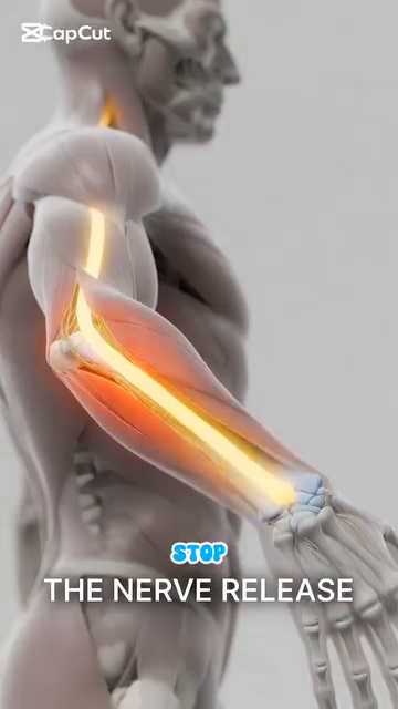 (Figure 1, Anatomy_Tay_Than_Kinh_Full.docx)
Figure 3 — STOP STRETCHING Warning
The critical message: stop passive stretching. Do nerve flossing instead.
Figure 5 — Nerve Flossing Position
Shows the alternating wrist + elbow motion: extend elbow + flex wrist ↔ flex elbow + extend wrist.
Figure 7 — Open Hand Skeleton — 8 Carpals as Mobile Base
Shows all 27 bones of the hand: 8 carpals, 5 metacarpals, 14 phalanges.
Figure 8 — Carpal Tunnel Cross-Section
Shows the 9 flexor tendons + median nerve packed into the 2 cm² tunnel.
Figure 9 — Fist — Thenar Muscle Group
Shows the thumb opposition mechanism. The thenar eminence = 50% of hand function.
Figure 10 — Extensor Tendon to Index Finger — Independent Control
The index finger has INDEPENDENT extensor control — important for delicate racquet face adjustment.
Figures 11–20 — Biceps, Triceps, Forearm Muscles (Tennis Anatomy Ch.3)
| Figure | Description | Image |
|---|---|---|
| 11 | Biceps brachii, brachialis, brachioradialis | DD3_arms_wrists_hands_01.png (Tennis Anatomy Fig.3.1) |
| 12 | Triceps brachii (3 heads) | DD3_arms_wrists_hands_02.png (Fig.3.2) |
| 13 | Forearm muscles inside | DD3_arms_wrists_hands_03.png (Fig.3.3a) |
| 14 | Forearm muscles outside | DD3_arms_wrists_hands_04.png (Fig.3.3b) |
| 15 | Triceps Cable Push-Down | DD3_arms_wrists_hands_05.png (Fig.3.4) |
| 16 | Triceps Push-Down — end position | DD3_arms_wrists_hands_06.png (Fig.3.5) |
| 17 | Triceps Rope Push-Down variation | DD3_arms_wrists_hands_07.png (Fig.3.6) |
| 18 | Half Dip — start position | DD3_arms_wrists_hands_08.png (Fig.3.7) |
| 19 | Half Dip — bottom position | DD3_arms_wrists_hands_09.png (Fig.3.8) |
| 20 | Wrist Curl — forearm flexor work | DD3_arms_wrists_hands_10.png (Fig.3.9) |
All image filenames verified to exist in Anatomy_Lab/images/DD3_arms_wrists_hands/.
🔗 CROSS-REFERENCES
| Topic in DD3 | See Also |
|---|---|
| Tennis elbow from over-grip | DD2 Shoulders — cuff vs deltoid imbalance, overuse patterns |
| Cubital tunnel / ulnar nerve | DD4 Trunk & Spine — C8-T1 nerve roots, neck posture |
| Wrist flexion / extension in stroke | DD1 Player in Motion — wrist angle at CONTACT (0–20° vs LOADED 90–110°) |
| Carpal tunnel in tennis players | DD8 Control System — proprioception decline, grip modulation |
| Triceps in serve | DD2 Shoulders — triceps long head crosses the shoulder, deceleration |
| Forearm muscles in pronation/supination | DD1 Player in Motion — racquet face control at contact |
📚 SOURCES
| Source | Type | What It Contributed |
|---|---|---|
Human anatomy/Anatomy_Tay_Than_Kinh_Full.docx |
User's Vietnamese notes (10 images) | Ulnar nerve pathway, cubital tunnel narrowing 55%, STOP STRETCHING message, nerve flossing technique, 27 hand bones, 8 carpals in 2 rows, carpal tunnel 2 cm², 3/10 to 7/10 grip rule, tendon gliding sequence |
Tennis Knowledge/7.Tennis Books in pdf/Tennis Anatomy ( PDFDrive ).pdf Ch.3 |
Reference textbook | Biceps/triceps/forearm anatomy, triceps push-down exercises, half dip, wrist curls |
Tennis Knowledge/7.Tennis Books in pdf/Tennis Anatomy ( PDFDrive ).pdf Ch.10 |
Same PDF | Tennis elbow, carpal tunnel syndrome in tennis players |
| Finestone & Rabinovitch, "Tennis elbow no more," Canadian Family Physician (2008), via tenniskb.github.io/tenniskb research report | Peer-reviewed reference | ECRB origin mechanism, "leading elbow" fallacy, elite-vs-recreational wrist angle data, dual-position (90°/180°) strengthening protocol |
End of DD3 — Arms, Wrists & Hands
Next: DD4 — Trunk & Spine (L4-L5, Piriformis, 3-Layer Back, Hip Hinge)