DD7 — Ankles & Feet
26 Bones, 33 Joints, the Windlass Mechanism, and Why Your Footwear Matters More Than Your Racket
📋 DOCUMENT MAP
The foot is a 26-bone, 33-joint, 19-muscle, 7,000+ nerve ending machine. It is the only part of your body in constant contact with the ground. It is the foundation of the kinetic chain (DD1). Get the foot wrong and nothing above it works right.
What it covers: the 26 bones in 3 zones (hindfoot, midfoot, forefoot), the windlass mechanism (the plantar fascia as a tension cable), the intrinsic vs extrinsic muscles, the 7,000 nerve endings and 30-millisecond reflex, the outside-leg bridge foot position (30–45°), and how tennis shoes affect (or destroy) foot function.
What it does NOT cover: the knee (DD6), the ankle joint in detail with ligament injuries (Tennis Anatomy Ch.10), or running shoe selection (beyond the tennis-specific note).
Reading time: 35–45 minutes.
📑 TABLE OF CONTENTS
| # | English |
|---|---|
| 1 | The 26 Bones in 3 Zones |
| 2 | The 19 Muscles — Intrinsic vs Extrinsic |
| 3 | The Plantar Fascia and the Windlass Mechanism |
| 4 | The Arches — Longitudinal and Transverse |
| 5 | The 7,000 Nerve Endings — The 30-ms Foot Reflex |
| 6 | The Outside-Leg Bridge — Foot Position at 30–45° |
| 7 | The Happy Feet Cue — Pre-Tension the Plantar Fascia |
| 8 | The Tennis Shoe Trap — 10 mm Heel Lift, 15 mm Toe Box |
Chapter 1 — The 26 Bones in 3 Zones
The human foot has 26 bones, organized in 3 functional zones: hindfoot (ankle area), midfoot (arch), forefoot (toes and metatarsals). Together they form 33 joints. The foot is NOT a rigid platform. It's an ADAPTIVE structure that changes shape with every step.
The 3 Zones of the Foot
| Zone | # Bones | Names | Function | Tennis Translation |
|---|---|---|---|---|
| Hindfoot | 7 | Talus, calcaneus (heel), navicular, cuboid + 3 cuneiforms (medial, intermediate, lateral) | Shock absorption (heel strike), talus pivot for inversion/eversion | The "spring" — heel strikes first on landing, then the talus pivots |
| Midfoot | 5 (navicular, cuboid, 3 cuneiforms) | Forms the ARCH — the bridge of the foot | Stores and releases elastic energy during gait | The "spring" itself. Calcaneus → metatarsals via plantar fascia. |
| Forefoot | 14 | 5 metatarsals + 14 phalanges (2 in big toe, 3 in each other toe) | Push-off during gait. The big toe is the LAST bone to leave the ground. | The "lever" — the great toe is critical for push-off. |
The 33 Joints — Why Mobility Matters
| Joint Type | # in Foot | Role |
|---|---|---|
| Hindfoot | 4 (subtalar, talocalcaneal, calcaneocuboid, talonavicular) | Inversion/eversion, plantar/dorsiflexion |
| Midfoot | ~9 (cuneonavicular, intercuneiform, cuneocuboid) | Arch support, fine adaptation |
| Forefoot | ~20 (tarsometatarsal, metatarsophalangeal, interphalangeal) | Push-off, gripping |
The Hindfoot — The Pivot Point
| Bone | Anatomy | Tennis Role |
|---|---|---|
| Talus | The "ankle bone" — sits between tibia (above) and calcaneus (below). No muscle attachment. Just a pivot. | Transmits forces from leg to foot. Any dysfunction → whole leg compensates. |
| Calcaneus (heel bone) | The largest foot bone. Achilles tendon attaches here. | The first bone to hit the ground on a step. |
| Navicular | Medial midfoot. Tibialis posterior attaches here. | The "keystone" of the medial longitudinal arch. |
| Cuboid | Lateral midfoot. Peroneus longus tendon passes through here. | Helps stabilize the lateral column during push-off. |
Source: Giai_phau_Ban_chan_Tennis.docx Ch.1-2 (Hệ xương và khớp). Tennis Anatomy Ch.7.
Chapter 2 — The 19 Muscles (Intrinsic vs Extrinsic)
The foot has TWO sets of muscles: INTRINSIC muscles (11 small muscles entirely within the foot) and EXTRINSIC muscles (8 muscles originating in the leg and inserting into the foot). They have different jobs.
The user's source makes the distinction: "Cơ chày trước, cơ mác dài, cơ tam đầu cẳng chân bám vào bàn chân từ xa. Chúng tạo lực lớn nhưng phản ứng chậm." (Tibialis anterior, peroneus longus, gastrocnemius attach to the foot from afar. They create large force but react slowly.)
The 19 Foot Muscles — Intrinsic vs Extrinsic
| Type | # | Examples | Primary Role | Tennis Translation |
|---|---|---|---|---|
| Intrinsic | 11 | Abductor hallucis, flexor digitorum brevis, quadratus plantae, lumbricals (4), interossei (4 — but only 3 in foot), adductor hallucis | Foot shape, fine control, proprioception | Adjust foot to surface. Hold arch during push-off. |
| Extrinsic | 8 | Tibialis anterior, tibialis posterior, peroneus longus, peroneus brevis, peroneus tertius, extensor digitorum longus, extensor hallucis longus, flexor digitorum longus | Force production, gross movement | Push-off, lateral stability, ankle positioning |
The Intrinsic Muscles — The Forgotten Heroes
The intrinsic foot muscles do NOT produce large force. They produce FINE CONTROL. They adjust the foot's shape to the ground. They maintain the arch during dynamic loading. They fire 30 milliseconds BEFORE the extrinsic muscles — pre-activating the foot for the upcoming load.
The 50+ truth: the intrinsic foot muscles atrophy from shoe wearing. Modern shoes SUPPORT the arch, so the intrinsic muscles don't need to work. They atrophy. By age 60, the intrinsic foot muscle mass is ~30% less than at age 20.
The fix: barefoot time. 5–10 minutes a day on grass or carpet. Walk, stand, do "short foot" exercises (try to shorten the foot without curling the toes). After 4 weeks, intrinsic strength returns.
Source: Giai_phau_Ban_chan_Tennis.docx Ch.3-4.
Chapter 3 — The Plantar Fascia and the Windlass Mechanism
The plantar fascia is a thick band of connective tissue that runs from the calcaneus (heel) to the bases of the toes. It's like a CABLE running under the foot. When the big toe extends (during push-off), the cable tightens. This tightens the arch. This is the WINDLASS mechanism — the foot becomes a rigid lever for push-off.
The windlass is the reason you can SPRINT. Without it, the foot would collapse on push-off. With it, the foot becomes a rigid lever — pushing 2–3× more efficiently.
The windlass for tennis: every split-step, every push-off, every wide recovery — the windlass activates. A foot with poor windlass (collapsed arch, restricted toe extension) loses 20–30% of push-off power. The knee compensates → patellar tendonitis. The hip compensates → adductor strain.
The Windlass Mechanism — Step by Step
| Step | Position | What Happens |
|---|---|---|
| 1. Foot flat | Heel strike, foot in contact with ground | Windlass is LOOSE. Foot is MOBILE. Adapts to surface. |
| 2. Midstance | Body weight over foot, arch compresses 3–4 mm | Windlass still loose. Intrinsic muscles fire. |
| 3. Heel off | Heel lifts, weight shifts to forefoot | Plantar fascia starts to tension. |
| 4. Big toe extension | Big toe dorsiflexes (extension) 60–90° as body moves over it | WINDSLASS ACTIVATES. Cable tightens. Arch rises. Foot becomes rigid. |
| 5. Push-off | Foot is rigid lever. Toe-off. | Maximum push-off power. |
The 3 Failures of the Windlass
| # | Failure | What Happens | Tennis Result |
|---|---|---|---|
| 1 | Hallux rigidus (big toe arthritis) | Big toe can't extend 60°. Windlass doesn't engage. | Loss of push-off power. Knee compensation. |
| 2 | Plantar fasciitis | Plantar fascia inflamed. Cable is "too tight" already. | Pain with first step in morning. Micro-tears in fascia. |
| 3 | Posterior tibial tendon dysfunction | The muscle supporting the arch is weak. Arch collapses during midstance. | "Adult-acquired flat foot." Windlass can't engage. |
The 50+ Windlass Reality — Why Push-Off Power Drops
By age 60, big toe extension typically decreases by 15–25°. The windlass engages less effectively. Push-off power drops 20–30%.
The fix: big toe mobility exercises. Sit with foot on opposite knee. Pull big toe back (extension) gently. Hold 30 sec. 5 reps × 3/day. After 4 weeks, extension improves 10–15°.
The tennis cue: "press through the big toe." When you push off for a forehand, the LAST thing touching the ground should be the big toe pad. If the push-off is on the ball of the foot or the small toes, the windlass isn't engaging.
Source: Giai_phau_Ban_chan_Tennis.docx Ch.5 (Cân gan chân và cơ chế windlass).
Chapter 4 — The Arches (Longitudinal and Transverse)
The foot has TWO arches that work together: the MEDIAL LONGITUDINAL ARCH (the inside of the foot, from heel to big toe) and the TRANSVERSE ARCH (across the foot, between the metatarsals). Together they create the spring and the strut.
The medial longitudinal arch: the more famous one. It absorbs shock during heel strike and rebounds during push-off. Supported by the plantar fascia, tibialis posterior tendon, spring ligament, and intrinsic muscles.
The transverse arch: the unsung hero. It runs across the foot at the level of the cuneiforms and metatarsal bases. It prevents the foot from splaying during push-off. In tennis, the OUTSIDE LEG uses the transverse arch as a STRUT — the foot is angled 30–45°, and the transverse arch converts horizontal force into vertical compression.
The 2 Arches — Different Roles
| Arch | Shape | Function | Tennis Role |
|---|---|---|---|
| Medial longitudinal | Curves from calcaneus to 1st metatarsal head (big toe) | Shock absorption (heel strike), elastic recoil (push-off) | "Spring" — stores and releases energy |
| Transverse | Curves across the cuneiforms + metatarsal bases | Prevents splay, provides lateral rigidity | "Strut" — converts horizontal force into vertical compression in wider stance |
The Arch Collapse Chain (Pronation → Valgus → Knee)
When the medial longitudinal arch collapses (over-pronation): the foot rolls inward. The tibia rotates internally. The knee goes into valgus. The hip drops.
The kinetic chain reaction: foot → tibia → knee → hip → lumbar. Each link rotates inward. The chain ends at L4-L5 (DD4).
The tennis trap: players with collapsed arches feel "slower." They're not slower in the legs. They're losing force in the chain. The arch absorbs the force that should go to the ball.
The "Short Foot" Exercise — Arches Activation
| Step | Action | Why |
|---|---|---|
| 1. Stand barefoot, weight even | Base position | Avoid compensation |
| 2. Try to shorten the foot (pull ball of foot toward heel) WITHOUT curling toes | The KEY movement | Curls = extrinsic. Shortening = intrinsic. |
| 3. Hold 10 seconds | Endurance | Build intrinsic muscle endurance |
| 4. 10 reps × 3/day | Frequency | Atrophy reversal takes 4–6 weeks |
Source: Giai_phau_Ban_chan_Tennis.docx Ch.6.
Chapter 5 — The 7,000 Nerve Endings (The 30-ms Foot Reflex)
The sole of the foot has more than 7,000 nerve endings. They are the body's first line of information about the world. They detect pressure, vibration, temperature, pain. They send signals UP the spinal cord in 30 milliseconds — FASTER than conscious awareness (~80 ms).
The 30-ms reflex: when the foot slips on a wet court, the nerve endings fire. The signal reaches the spinal cord in 30 ms. The spinal cord sends a CORRECTION back to the muscles in another 30 ms. Total response: 60 ms. You have already braced yourself BEFORE you consciously know you slipped.
The 50+ decline: by age 65, the number of nerve endings in the sole decreases by ~30%. Proprioception declines. The 30-ms reflex becomes 50-ms reflex. Recovery time from a slip increases. The risk of falling increases.
The Pressure Distribution Map
| Pressure Zone | Pro Player | 3.5 Club Player | Why It Matters |
|---|---|---|---|
| Big toe (hallux) | 28% | 5–10% | Big toe is the FINAL lever in push-off. Pros use it. |
| Pinky toe (5th metatarsal) | 22% | 5–10% | Lateral stability. Pros use it. |
| Other toes + forefoot | 35% | 40–45% | Spread evenly. |
| Midfoot | 10% | 25–35% | The 3.5 player has FLAT midfoot contact (arch collapse). |
| Heel | 5% | 15–20% | Heel too loaded = no push-off. |
The Tennis Foot Reflex Test — A Self-Check
Stand on one leg, eyes closed. Have a friend time you.
Pro standard: 30+ seconds, no wobble.
50+ standard: 10–15 seconds is normal. Below 10 seconds = high fall risk.
The fix: practice daily. Eyes-closed standing on one leg is the only exercise that has been shown to REDUCE fall risk in 50+ adults. 3×30 sec each side, daily.
Source: Giai_phau_Ban_chan_Tennis.docx Ch.7-8 (7,000 nerve endings, 30 ms reflex, pressure distribution).
Chapter 6 — The Outside-Leg Bridge (Foot Position at 30–45°)
When you hit a forehand in open stance, your OUTSIDE LEG (front leg, left for right-hander) is the BRIDGE. It supports up to 3–5× body weight in compressive load. The foot MUST be angled 30–45° (not straight).
Why 30–45°? the angle uses the TRANSVERSE ARCH as a STRUT. Horizontal force from the swing decomposes into vertical compression. The force travels up through the talus, tibia, knee, and into the femur — all in a near-straight line.
If the foot is straight (0°): all the horizontal force becomes valgus stress on the knee → medial meniscus load → IT band friction. The knee hurts. The lateral foot collapses.
If the foot is angled >60° (over-rotated): the foot loses its strut function. The transverse arch collapses. The force becomes shear on the knee.
The Outside Leg Foot Position — 3 Zones
| Foot Angle | Zone | Force Decomposition | Knee Effect |
|---|---|---|---|
| 0° (straight) | Dangerous | All horizontal → valgus | High meniscus + IT band load |
| 30–45° | OPTIMAL | Horizontal → vertical compression | Low knee load. Maximum force transfer. |
| >60° | Over-rotated | Loss of strut function | Shear on knee. Arch collapse. |
The 3-Point Foot Anchor — Tripod Foot
The tripod foot is the GOLDEN position for groundstrokes. Three points of contact: heel (center), ball of foot under big toe, ball of foot under pinky toe. These three points form a triangle. The triangle is RIGID. The center of the triangle is the "tripod" — the load-bearing center.
The test: stand barefoot. Try to lift just the big toe while keeping the ball of foot under the pinky toe on the ground. If you can't, your tripod is collapsed. Practice "short foot" daily until you can.
Source: Giai_phau_Ban_chan_Tennis.docx Ch.9-11.
Chapter 7 — The Happy Feet Cue (Pre-Tension the Plantar Fascia)
"Happy feet" in tennis means the feet are constantly in motion — never flat on the ground for long. The user defines it more specifically: "Giữ gót lơ lửng giúp cân gan chân ở trạng thái tiền căng." (Keeping the heel floating keeps the plantar fascia in pre-tension.)
The pre-tension principle: when the plantar fascia is pre-tensioned (heel off ground), the foot is ready to spring into action. The split-step can launch in any direction instantly. No delay. No "loading time."
The Happy Feet Position — Anatomy
| Element | Position | Muscle State |
|---|---|---|
| Heel | Slightly off ground (1–2 cm) | Calf (gastrocnemius + soleus) lightly contracted |
| Ball of foot | In contact with ground | Tibialis anterior relaxed |
| Big toe | Light contact, not gripping | Flexor hallucis longus relaxed |
| Plantar fascia | PRE-TENSIONED | Ready to spring |
| Arch | Slightly raised (active) | Intrinsic muscles engaged |
The Cost of Flat Feet in Tennis
If your heel stays flat on the ground for >2 seconds during a point, you have "lazy feet." The plantar fascia goes slack. The split-step reaction time increases from 100 ms to 250 ms. You lose 150 ms — enough for an opponent to hit a winner.
The 50+ habit: most recreational players let the heel drop between shots. They're waiting for the next ball. They're flat-footed. The pro keeps the heel up — always ready. The pro's split-step is half the time of the recreational player.
The fix: the "elevator heel." Between every shot, lift the heel 1 cm. Count: split-step → hit → recover → HEEL UP. Make it a rhythm. After 2 weeks of conscious practice, it becomes automatic.
Source: Giai_phau_Ban_chan_Tennis.docx Ch.10 (Liên hệ với happy feet).
Chapter 8 — The Tennis Shoe Trap (10 mm Heel Lift, 15 mm Toe Box)
The user's source has a SHOCKING statistic: "Hầu hết giày tennis nâng gót 10 mm, thu hẹp mũi 15 mm so với bàn chân tự nhiên. Hệ quả là ngón cái không duỗi được, windlass giảm hiệu quả, cơ nội sinh teo dần." (Most tennis shoes lift the heel 10 mm and narrow the toe box by 15 mm compared to the natural foot. The result is the big toe can't extend, the windlass is less effective, and intrinsic muscles atrophy over time.)
The 10 mm heel lift shortens the calf. Over time, the calf adapts to the shorter length. When you take the shoe off, the calf "feels" too long. You feel tight. You stretch the calf. The calf SHORTENS further. A vicious cycle.
The 15 mm toe box narrowing prevents the big toe from extending 60–90° at push-off. The windlass doesn't engage. Push-off power drops 20–30%.
The Tennis Shoe Numbers — What to Look For
| Number | What It Means | What You Want |
|---|---|---|
| Heel-to-toe drop | The mm of lift from heel to toe | 4–8 mm max. NOT 10+ mm. |
| Toe box width | The mm of horizontal space for toes | Your natural foot width + 5–10 mm. NOT -15 mm. |
| Heel counter stiffness | How rigid the back of the shoe is | Moderate. Not floppy, not board-stiff. |
| Midsole cushioning | How much shock absorption under the heel | Moderate. Too soft = unstable. Too hard = joint impact. |
| Outsole pattern | The tread pattern on the bottom | Herringbone for hard courts. Modified herringbone for clay. |
| Upper material | The fabric on top | Breathable mesh. Avoid heavy leather (waterlogging). |
The 3 Tennis Shoe Mistakes
| # | Mistake | Consequence | The Fix |
|---|---|---|---|
| 1 | Wearing shoes >6 months old | Midsole cushioning degrades 30–50% | Replace every 4–6 months for regular players |
| 2 | Wearing the wrong surface shoe | Wrong tread = slipping or sticking | Hard court shoes on hard court. Clay shoes on clay. |
| 3 | Tight toe box "for support" | Big toe can't extend. Windlass fails. | Buy shoes with TOE ROOM. Wiggle toes inside. |
The 50+ Foot Longevity Protocol
| Element | Frequency | Why |
|---|---|---|
| Barefoot time on grass or carpet | 5–10 min/day | Re-activates intrinsic foot muscles |
| Short foot exercise | 3×10/day | Reverses arch collapse |
| Big toe extension stretch | 5×30 sec × 3/day | Maintains windlass engagement |
| Single-leg balance eyes closed | 3×30 sec × 3/day | Maintains 7,000 nerve endings |
| Replace tennis shoes every 4–6 months | Per pair | Midsole degrades |
| Rotate 2 pairs | If possible | Midsole recovers between sessions |
Source: Giai_phau_Ban_chan_Tennis.docx Ch.12-14. Reference: minimalist shoe research, foot health literature.
📋 DD7 CARD — Printable
The foot is a 26-bone, 33-joint, 7,000-nerve-ending machine. The windlass (plantar fascia as cable) makes push-off powerful. Outside-leg bridge: foot at 30–45°. Tennis shoes with 10 mm heel lift + 15 mm toe box narrowing destroy foot function over time.
- 26 bones / 33 joints / 19 intrinsic+extrinsic muscles
- 7,000+ nerve endings in sole
- 30 ms reflex (foot → spinal cord → muscle)
- Big toe extends 60–90° for windlass to engage
- Push-off power drops 20–30% without windlass
- Tennis shoes: 10 mm heel lift, 15 mm toe box narrowing
- Pros: 28% pressure on big toe, 22% on pinky
- 3.5 players: 5–10% on big toe, 25–35% on midfoot
Letting the heel drop flat between shots (lazy feet). The plantar fascia goes slack. The split-step reaction time goes from 100 ms to 250 ms — enough time for the opponent to hit a winner. The fix: "Elevator heel" — keep the heel 1 cm off the ground.
- Short Foot: stand barefoot, shorten foot without curling toes. 10 reps × 3/day.
- Big Toe Extension Stretch: pull big toe back, hold 30 sec. 5 reps × 3/day.
- Single-Leg Balance Eyes Closed: 30 sec each side. 3 reps × 3/day. Reduces fall risk in 50+.
"Heels up, tripod foot, press through the big toe."
🖼️ ILLUSTRATIONS
38 images available in Anatomy_Lab/images/DD7_ankles_feet/ (19 from Giai_phau_Ban_chan_Tennis.docx + 19 from Tennis Anatomy PDF).
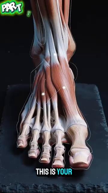
Figure 2–3 — Muscle-Tendon Structure of Dorsum of Foot
| Figure | Description | Image |
|---|---|---|
| 2 | Muscle-tendon structure during heel-lift preparation | Giai_phau_Ban_chan_Tennis__img02.jpg |
| 3 | Same — different angle | Giai_phau_Ban_chan_Tennis__img03.jpg |
Figure 4–7 — Windlass Mechanism
| Figure | Description | Image |
|---|---|---|
| 4 | Forefoot deformation under body weight (windlass mechanism) | Giai_phau_Ban_chan_Tennis__img04.jpg |
| 5 | Same — different view | Giai_phau_Ban_chan_Tennis__img05.jpg |
| 6 | Same — different view | Giai_phau_Ban_chan_Tennis__img06.jpg |
| 7 | Same — different view | Giai_phau_Ban_chan_Tennis__img07.jpg |
Figure 8–11 — Sole Nerve Endings
| Figure | Description | Image |
|---|---|---|
| 8 | Nerve endings of sole and orientation role | Giai_phau_Ban_chan_Tennis__img08.jpg |
| 9 | Same — different angle | Giai_phau_Ban_chan_Tennis__img09.jpg |
| 10 | Same — different angle | Giai_phau_Ban_chan_Tennis__img10.jpg |
| 11 | Same — different angle | Giai_phau_Ban_chan_Tennis__img11.jpg |
Figure 12–14 — Pressure Distribution
| Figure | Description | Image |
|---|---|---|
| 12 | Pressure control when heel is fully lifted | Giai_phau_Ban_chan_Tennis__img12.jpg |
| 13 | Same — different view | Giai_phau_Ban_chan_Tennis__img13.jpg |
| 14 | Same — different view | Giai_phau_Ban_chan_Tennis__img14.jpg |
Figure 15–19 — Outside-Leg Bridge & Alcaraz Comparison
| Figure | Description | Image |
|---|---|---|
| 15 | Alcaraz mid-run, foot not yet tripod | Giai_phau_Ban_chan_Tennis__img15.jpg |
| 16 | Same — different view | Giai_phau_Ban_chan_Tennis__img16.jpg |
| 17 | Same — different view | Giai_phau_Ban_chan_Tennis__img17.jpg |
| 18 | Recovery + outside-leg bridge creation | Giai_phau_Ban_chan_Tennis__img18.jpg |
| 19 | Same — different view | Giai_phau_Ban_chan_Tennis__img19.jpg |
Figures 20–38 — Tennis Anatomy PDF Ch.7 (Foot/Ankle)
| Figure | Description | Image |
|---|---|---|
| 20 | Ankle joint bones | DD7_ankles_feet_01.jpg (Tennis Anatomy Fig.7.x) |
| 21 | Subtalar joint detail | DD7_ankles_feet_02.jpg |
| 22 | Achilles tendon attachment | DD7_ankles_feet_03.jpg |
| 23 | Plantar fascia anatomy | DD7_ankles_feet_04.jpg |
| 24 | Tibialis posterior tendon | DD7_ankles_feet_05.jpg |
| 25–38 | Various foot/ankle exercise demonstrations | DD7_ankles_feet_06.jpg through _19.jpg |
All image filenames verified to exist in Anatomy_Lab/images/DD7_ankles_feet/.
🔗 CROSS-REFERENCES
| Topic in DD7 | See Also |
|---|---|
| Foot tripod for groundstrokes | DD1 Player in Motion — the 6 critical angles at contact |
| Outside-leg bridge 30–45° | DD6 Knees — knee over 2nd toe, foot under hip |
| 7,000 nerve endings and proprioception | DD8 Control System — vestibular + vision + proprioception |
| Arch collapse → valgus → knee | DD6 Knees — ACL mechanism, patellofemoral pain |
| Windlass for push-off | DD1 Player in Motion — kinetic chain, ground reaction force |
| Happy feet and split-step | DD8 Control System — reaction time, vision |
| Tennis shoe selection | DD6 Knees — joint preservation, less force on cartilage |
📚 SOURCES
| Source | Type | What It Contributed |
|---|---|---|
Human anatomy/Giai_phau_Ban_chan_Tennis.docx |
User's Vietnamese notes (19 images) | 26 bones 33 joints, hindfoot/midfoot/forefoot, intrinsic vs extrinsic muscles, windlass, longitudinal + transverse arches, 7,000 nerve endings 30 ms reflex, pressure distribution 28%/22%, outside leg bridge 30–45°, happy feet, tennis shoe trap 10 mm/15 mm, rehabilitation program |
Tennis Knowledge/7.Tennis Books in pdf/Tennis Anatomy ( PDFDrive ).pdf Ch.7 (Legs) |
Reference textbook | Foot anatomy, ankle joint, Achilles tendon, calf muscles |
End of DD7 — Ankles & Feet
Next: DD8 — The Control System (Vestibular, Proprioception, Vision, 50+ Body)